Nuclear medicine uses radioactive material (usually in the form of a powder or liquid) to diagnose and treat diseases.
Several different techniques are used to investigate or treat a wide range of conditions.
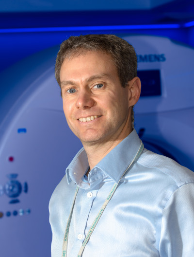
The nuclear medicine team is a multidisciplinary team and includes doctors, technologists, radiopharmacists, physicists, radiographers, healthcare assistants and administrative staff.
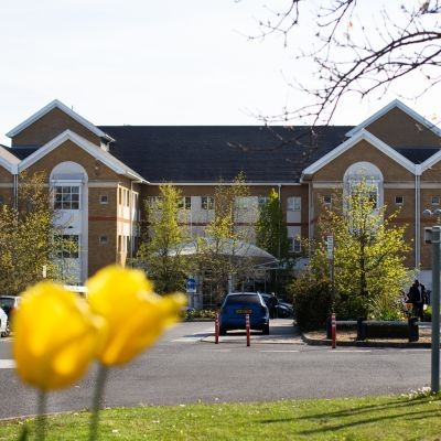
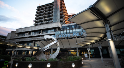
Our nuclear medicine services are based at the Royal Free Hospital and Barnet Hospital.
Gamma camera scanning involves the injection of a radioactive chemical into the body, which gives off radioactivity called gamma rays. These are then picked up on a scan.
The technique is used to examine how your body metabolises the chemical.
This is a highly versatile technique and can be used to examine a wide range of medical conditions.
Sometimes it takes time for the chemical to be properly distributed in the body before the scan can be done. This could be up to three or four hours. You do not need to stay in the nuclear medicine department during this time.
The type and duration of scan depends on the type of investigation, but varies between 15 minutes to over one hour. You must try to remain still during the scan.
PET/CT (positron emission tomography/computerised tomography) is an imaging technique that produces 3D images of a radioactive chemical distributed in the body.
You will receive an injection of a chemical into a vein before your scan.
The scan typically takes between 20 to 30 minutes. During the scan, the bed will slowly move through the scanner. You must stay as still as possible.
You must leave all jewellery at home if possible (rings can be kept on if preferred). Wearing jewellery during PET/CT scans is not a safety concern, but it can affect the quality of the images.
There are different types of PET/CT scans and you will be given instructions on how to prepare for your scan, depending on the type you are having. For example, you may need to fast for a set period of time before your scan and avoid vigorous physical exercise.
Your consultant or clinical nurse specialist will provide you with information about how to prepare for your particular scan.
There is a video by London Cancer that provides more information about PET/CT scans.
Radionuclide therapy uses radioactive material which emits short-range radiation in order to treat disease. Different treatment techniques are used in order to treat different diseases.
How long you need to spend in hospital will depend on the type of therapy you are having.
As some of these therapies emit some longer-range radiation, which other people could be exposed to, you will be given advice about how to minimise exposure to other people.
GFR is a measure of how well your kidneys are functioning.
The technique used in the nuclear medicine department is more accurate than standard estimated GFR (eGFR), which is done via a simple blood test.
The technique involves administering a very small quantity of a radiotracer into one of your veins. A blood sample is taken from a different vein.
The amount of radiotracer remaining in the blood can then be accurately determined, which shows how quickly the blood is being filtered by the kidneys.
DEXA, or DXA, is a technique to measure bone mineral density (BMD).
During the scan, you will need to lie down while the DEXA scanner arm moves over your body, taking several images using X-rays.
Two different X-rays are used, which makes it possible to determine the amount of X-rays absorbed by bone, versus the amount absorbed by other tissue, which provides information about BMD.
Patients are mostly referred to the nuclear medicine department from other healthcare professionals or teams within the hospital.
GP referrals are accepted for DEXA.
 Translate


