Translate
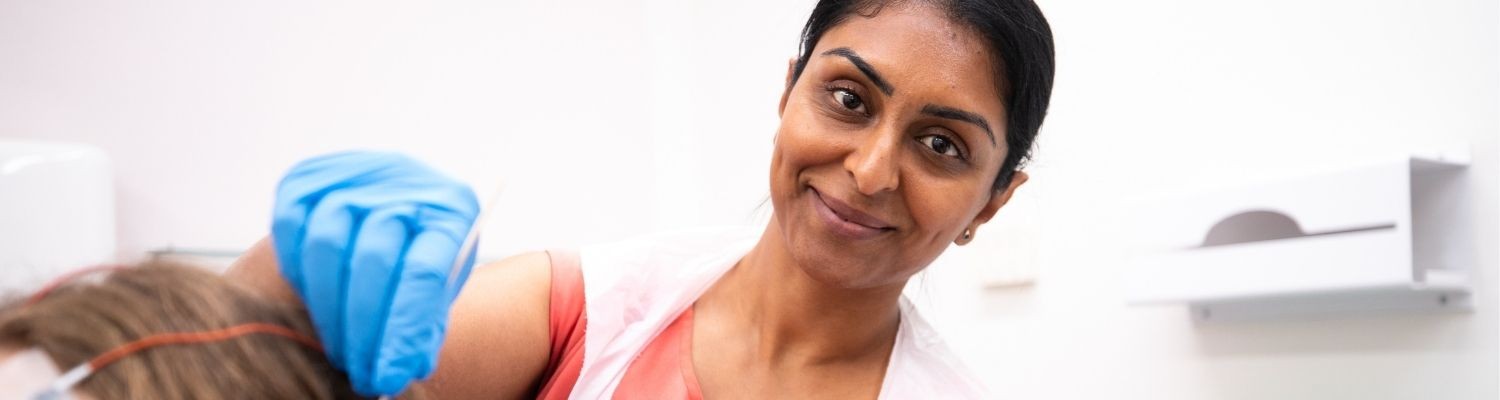
Our neuroscience service deals with conditions affecting the brain, eyes, speech, swallowing and limbs.
For example, a neurologist may treat multiple sclerosis (MS), Parkinson's disease, epilepsy and patients who have had a stroke.
A neurologist could also treat Tourette's syndrome and Alzheimer's disease, among other medical conditions.
Our nervous system treatment and neurological tests include:
- neurology
- neuropsychology, looking at brain function and brain problems
- neurophysiology, providing treatment for nervous system disorders and muscle diseases
Patients will be treated within the department or referred to one of our speciality services.
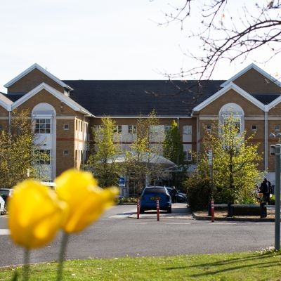
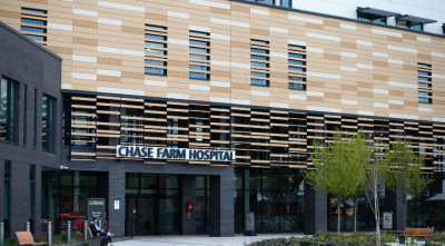
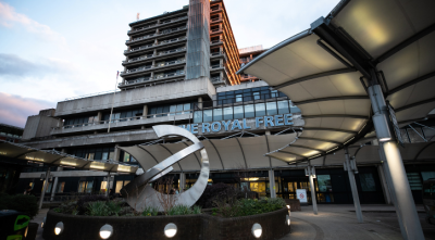
Clinics are run at the Royal Free Hospital, Barnet Hospital and Chase Farm Hospital, as well as Edgware Community Hospital and Potters Bar Community Hospital.
Our neurology department deals with several medical conditions including epilepsy, Parkinson’s disease and MS at Barnet Hospital, Chase Farm Hospital, and the Royal Free Hospital.
We have over 20 neurologists running more than 30 clinics per week and seeing more than 1,000 patients a year. Our team also manages about 300 patients who have had a stroke.
Our neurology department leads in education, providing undergraduate and postgraduate teaching and medical research.
Alongside our general neurology clinics, the team specialise in:
- botulinum toxin injections
- epilepsy
- motor neurone disease
- multiple sclerosis
- neuro-ophthalmology
- neurosarcoidosis
- Parkinson’s disease
- peripheral neuropathy
They also specialise in medical treatment related to cognitive neurology, headaches, neuro-genetics and HIV neurology, among others.
Our epilepsy service at the Royal Free Hospital aims to enhance the quality of life for those diagnosed with epilepsy and their families.
We do this through treatment, support, provision of advice and education regarding epilepsy.
The epilepsy service also aims to increase levels of understanding on a wide range of issues related to epilepsy, reduce relapses and side-effects where treatment is necessary, and aid the reduction in misdiagnosis.
Reports combined with formal assessments enable the service to continue running several outpatient clinics per week and assess, plan and treat others as inpatients when necessary.
Clinical nurse specialist
The clinical nurse specialist (CNS) for epilepsy will coordinate and liaise with other healthcare professionals in accordance with National Institute for Health and Clinical Excellence (NICE) guidelines.
In response to this, the CNS will see patients within nurse-led clinics twice per week, and will follow-up the care and treatment of others via formally arranged telephone consultations once per week.
The nurse will also aim to provide a level of training relevant to the needs of staff and carers.
The CNS primarily acts as a confidential point of contact for the majority of patients already known to the consultant neurologists and other specialists.
Patients may wish to focus on certain areas that concern them — these may include proposed or present treatment including side-effects, relapses, risk management, memory impairment, mood, maintaining continuity of services or accessing other services.
The nurse may also wish to raise other areas with the patients that would otherwise not necessarily be discussed. These may include:
- the possibility of sudden unexpected death in epilepsy (SUDEP) in certain patients
- reproductive issues including planning for pregnancy, fertility and sexual dysfunction
The multiple sclerosis clinical nurse specialist (MS CNS) service incorporates acute services at Chase Farm Hospital and the Royal Free Hospital, where the MS CNS’ are based, and commissioned services at the neurological rehabilitation centre at Edgware Community Hospital.
The MS CNS’ recognise the very individual needs of every person with MS through diagnosis and beyond. The service follows NICE guidelines and the National Service Framework for Long Term Conditions.
Their aim is to provide a resource for people with MS, which enables them to manage and respond appropriately to changes in their symptoms and enables them to achieve their best health and quality of life through support, education, information, medicine management and monitoring, signposting and referral.
Liaison with other healthcare professionals and members of the multidisciplinary team ensures timely specialist input to the variable and unpredictable symptoms associated with this condition.
Movement disorders are a group of neurological conditions that impact the way your body moves. The movement disorders service at Royal Free London offers an assessment for people with movement disorders including Parkinson’s disease, Parkinsonism, dystonia, tremor, tics, gait and balance disorders.
For general research enquiries and patient involvement opportunities please contact our patient and public involvement officer for Parkinson’s.
Email: rf.
Tel: 020 7794 0500 ext 36927
Movement disorder clinics
Outpatient movement disorder clinics are held at the Royal Free Hospital twice a week. The clinics are run by consultant neurologists and supported by clinical nurse specialists, the neurosciences team and the speech and language team.
Our health services for elderly people consultant, who has a specialist interest in Parkinson’s, runs a weekly clinic at the Royal Free Hospital and a Parkinson’s clinic is also held twice a month at Chase Farm Hospital.
If you are already taking medication, we always suggest you bring a list of all your medications, dosages and timings with you when you come to clinic. This is very useful for all the clinical staff to be able to help you quickly.
The neurophysiology department at the Royal Free Hospital is part of our neuroscience team, looking after nervous system disorders and muscle diseases.
It deals with epilepsy, patients with a trapped nerve, or nervous system disorders affecting the peripheral nervous system (not the brain or spinal cord).
Neurophysiology treatment: caring for the nervous system
The team monitor nervous system disorders and muscle diseases in a number of ways. These include:
- electroencephalography (EEG), which records brain activity in order to monitor brain function and brain problems
- electromyography (EMG) and nerve conduction studies, which look at electrical activity produced by skeletal muscles
- computer-controlled temperature testing, known as thermal thresholds, which investigate small fibre nerve dysfunction
Neurophysiology clinics: where do I go for treatment?
The neurophysiology department is on the 1st floor of the Royal Free Hospital, behind outpatient clinic 2. The reception is open from 9am to 5pm, Monday to Friday.
Contact details
- Tel: 020 7830 2072.
- Email: rfh-tr.
neurophysiology .@nhs.net
The neuropsychology department at the Royal Free Hospital is part of our neuroscience team and provides specialist assessments for people who have diagnosed or suspected neurological conditions who are under the care of a neurologist at the Royal Free London NHS Trust.
Neuropsychological assessments
A neuropsychological assessment helps to understand your thinking abilities. This is often referred to as cognitive abilities or skills. These abilities include memory, concentration, language, visual skills, and problem-solving skills.
The results can be used to:
- Help your neurologist make decisions about diagnosis or treatments that might help
- Monitor changes over time
- Provide advice on ways of coping with symptoms or difficulties in daily life
The neuropsychologist will ask you about any problems you have been having, when these started and whether they have changed over time, and what helps or makes the problems worse. They will also ask for details of your personal history, such as your education or work history, and about relevant physical and mental health problems during your life.
This is called a clinical interview’ It helps the neuropsychologist design the right assessment for your problems and interpret the findings correctly. This usually takes around 45 minutes.
You will have a face-to-face appointment with the neuropsychologist. They will use several pen-and-paper tasks to look at different thinking abilities / cognitive skills, explaining each task beforehand. This is called a ‘cognitive assessment’. Most people find it interesting to do. This appointment can last between two to three hours, with short breaks if needed.
In some cases, it is possible to complete both elements in one appointment. However, typically you would be offered two appointments, and occasionally an additional appointment will be needed.
After the appointment, the neuropsychologist will score your tests and send a report interpreting the findings to your doctor. This is usually your consultant but can be your neurologist. Depending on the purpose of your assessment, you and your GP may also receive a copy, or alternatively this can be shared with you once your neurologist has completed all your planned investigations.
Neuropsychology clinics: where do I go for treatment?
The neuropsychology department is on the Lower Ground Floor of The Grove, on the main Royal Free Hospital site. You will receive a map and information about how to find us with your appointment letter.
Contact us
- Tel: 020 7830 2292, extension 23089.
- Email: rf-tr.
neuropsychology .@nhs.net
Our friendly and approachable outpatient physiotherapy service at the Royal Free Hospital accepts referrals for all patients with neurological or vestibular conditions.
We offer:
- full assessment and individualised, evidence-based treatment
- assessment and advice during the early stages of neurodegenerative disease
- Parkinson’s disease early intervention clinic
- balance group
- self-management strategies
- education and empowerment
- health coaching
- health promotion
- provision and review of neurological splints and orthoses (braces)
- appropriate onward referral and signposting
We undertake vestibular rehabilitation for all peripheral and central vestibular disorders, including;
- BPPV
- labyrinthitis
- vestibular neuritis
- acoustic neuroma (vestibular schwannoma)
- Meniere’s disease and endolymphatic hydrops
- vestibular migraine
- visual vertigo
- secondary to vascular events or neurological disease
We undertake neurological rehabilitation for all neurological conditions, including:
- all stages of neurodegenerative disease e.g. MS, PD including early stages prior to significant signs/symptoms
- review of chronic neurological conditions such as stroke, traumatic brain injury, peripheral neuropathy, Guillain-Barré syndrome, chronic inflammatory demyelinating polyneuropathy, spinal cord injury
- functional movement disorders
- balance and falls
Referrals can be made by a consultant at the Royal Free Hospital or Royal National Throat, Nose and Ear Hospital, or by a GP. Please email all referrals.
If you have any queries, please email or call to discuss further.
Email: rf-tr.
Tel: 020 7794 0500 ext 34055 (please leave a message and we will return your call within one working day).
Parkinson's is a progressive neurological condition, which means that symptoms can often appear slowly and over a long time. Most people who develop Parkinson’s are aged 50 or over, but younger people can get it too. In the UK it is estimated that one person in every 500 has Parkinson’s, that’s about 127,000 people. There is currently no cure for Parkinson’s and we don’t yet know why people get the condition. However much research is taking place worldwide looking at all avenues of potential causes, which is helping us understand Parkinson’s better.
Symptoms
The main symptoms of Parkinson’s could be tremor, rigidity, slowness of movement or a combination of these. They are generally known as 'motor' symptoms, being to do with movement. As well as affecting movement, people with Parkinson’s may find that other issues, such as tiredness, pain, depression and constipation, can have an impact on their day to day lives. However, different people can experience the condition differently.
Treatment
Drug treatment is the main method used to control symptoms caused by the loss of dopamine. The main aim of these drugs is to increase the level of dopamine that reaches the brain and stimulate the parts of the brain where the dopamine works. There are many different drugs prescribed for Parkinson’s and research into treatments is improving the options all the time. Drugs are reviewed over time to make sure the best combination works well. Therapy such as physiotherapy, speech and language therapy, occupational therapy and diet can help in dealing with daily life. More information on treatments and therapies can be found on the Parkinson's UK website.
Parkinson’s service
Our service offers diagnostic assessment from a team of neurologists specialising in Parkinson’s and other movement disorders, which is supported by a Parkinson’s disease specialist nurse. This allows for clinical monitoring and medication adjustments and provides a continuous point of contact for information about clinical matters and other issues that concern people with Parkinson’s and their carers.
This service aims to enhance the quality of life for those diagnosed with Parkinson’s and their families by providing support, advice and education about Parkinson’s disease.
Neurosarcoidosis
Patients can be referred by their GP. Patients, their relatives and doctors should contact the sarcoidosis centre at the Royal Free Hospital for advice on how to refer or be referred for treatment, care, and research.
Neurological rehabilitation
Referrals can be made by a consultant at the Royal Free Hospital or Royal National Throat, Nose and Ear Hospital, or by a GP. Please email all referrals to rf-tr.