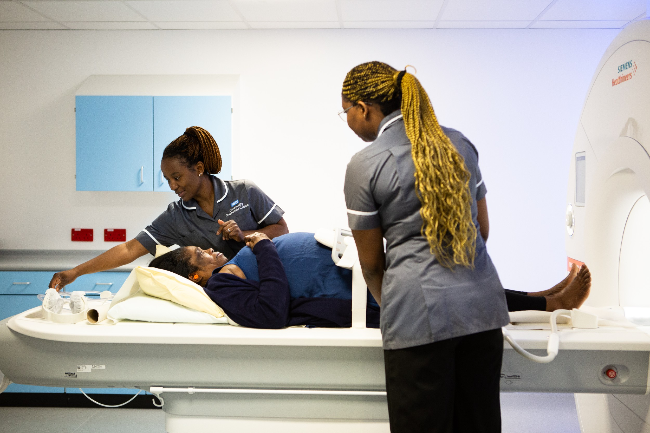
Our radiology service is a pioneer of innovation and excellence in the field of medical imaging.
A world-class facility, the service provides advanced and comprehensive diagnostic services to the community. The team provide formal reports on your images, which are sent to your referring doctor to guide any necessary treatment.
The service works across Barnet Hospital, Chase Farm Hospital, North Middlesex University Hospital and the Royal Free Hospital, providing easy accessibility and convenience for all our patients, supporting our emergency departments, inpatient and outpatient care, and GP referrals.
It provides precise and timely diagnostic information, and works to ensure every patient’s visit is comfortable, that they are fully informed, and that they receive unparalleled expertise.
Further down this page are the individual services within radiology, and what they mean.
Opening times
- Outpatient and general (walk-in service) X-ray: Monday to Friday, 9am to 4.30pm across all sites except for North Middlesex University Hospital which is open from 9am to 6pm, Monday to Friday
- Fluoroscopy appointments: Monday to Friday, 9am to 4.30pm
- Ultrasound appointments: Monday to Friday, 9am to 5pm across all sites except for North Middlesex University Hospital which is open from 9am to 7.30pm, Monday to Friday
- CT appointments: Monday to Friday, 8am to 6.30pm; Saturday, 9am to 5pm across all sites except for North Middlesex University Hospital which is open from 9am to 5pm, Monday to Friday
- MRI appointments: Monday to Sunday, 8am to 8pm across all sites except for North Middlesex University Hospital which is open from 8am to 9pm, Monday to Friday
- PET/nuclear medicine: Monday to Friday, 8am to 8pm across all sites except for North Middlesex University Hospital which is open from 9am to 5pm, Monday to Friday
- DEXA appointments: Monday to Friday, 8am to 8pm
- Emergency CT and X-ray: 24 hours a day, 7 days a week
Diagnostic radiographers are healthcare professionals trained to perform imaging examinations such as X-rays, MRI scans, CT scans, or nuclear medicine tests.
Those who perform ultrasound scans are referred to as sonographers.
Radiographers also support radiologists during interventional procedures and, in some cases, are trained to formally report X-rays.
Radiologists are medical doctors trained to interpret imaging. They provide reports on your images, which are then sent to the doctor who referred you.
Radiologists who perform procedures, such as biopsies or embolisation, are known as interventional radiologists.
Radiology nurses specialise in assisting radiologists during interventional procedures, such as biopsies or embolization.
They carefully monitor you before, during, and after your radiology procedure to ensure your safety and comfort.
An X-ray is a quick and non-invasive procedure that captures images of the internal structures of the body, aiding in the diagnosis of various bone and joint ailments.
This technology relies on a controlled source of X-rays, with the images displayed on a digital sensor and computer screen.
Ultrasound is a painless and non-invasive procedure that utilises high-frequency sound waves to create images of the internal structures of the body.
This technique is used extensively in monitoring pregnancies and diagnosing conditions involving organs such as the liver, kidneys and blood vessels.
A CT scan is a quick procedure that offers detailed two-dimensional or three-dimensional images of the body, helping in the precise diagnosis and management of diseases.
It sometimes requires the use of contrast (X-ray dye) to enhance image quality.
MRI scans use powerful magnets and radiowaves to generate detailed cross-sectional images of the body, providing insights often not available from other techniques.
This safe procedure can take longer than conventional X-rays or CT scans, and might not be suitable for people with certain medical devices such as pacemakers.
A dual-energy X-ray absorptiometry (DEXA) scan is a specialised X-ray procedure that measures bone mineral density, aiding in the diagnosis of osteoporosis and assisting in the planning of appropriate healthcare routines.
It is an essential tool in assessing the risk of osteoporosis, particularly in women aged over 50 and men aged over 60.
Nuclear medicine is a specialised area in healthcare that uses radioactive tracers, or radiopharmaceuticals, to evaluate how different parts of the body are functioning.
This method is instrumental in both diagnosing various diseases and guiding the right treatment plans, by providing clear images of bodily processes at a molecular level.
Fluoroscopy takes moving pictures of the internal structures of the body using a series of low-dose X-rays. This allows real-time imaging of organs and guided interventional procedures.
It is commonly used in angiography, venography and outlining body structures.
A PET scan is a unique imaging technique that utilises a special radioactive substance to detect metabolically active tissues, helping in the identification of tumours or inflammatory conditions.
This technique is known for its accuracy in pinpointing areas with increased metabolic activity
Mammography is a vital tool in women's health, utilising X-rays to detect irregularities or early signs of breast cancer.
Regular screenings can ensure early detection and effective management of breast cancer.
We provide a range of procedures at the Royal Free Hospital and Barnet Hospital for most areas of interventional radiology (IR), and we are a hub for IR in north central London.
The team is made up of radiologists, radiographers, nurses, and healthcare assistants, all working closely with our clinician colleagues.
What is IR?
IR is one of the most innovative and fastest-growing specialities in the field of medicine.
An interventional radiologist is a medical doctor who performs image-guided procedures, fully interpreting the imaging required to guide and monitor a patient’s response to those procedures.
Advances in medical imaging have allowed IR to perform the most complex of tasks through a small pinprick in the skin, advancing catheters, guidewires, or needles to targets deep inside the body, while avoiding damage to critical surrounding tissues.
Our procedure rooms contain imaging equipment that allows procedures to be performed in real-time, using X-rays, ultrasound and CT.
Because of this keyhole approach, IR procedures are often termed ‘minimally invasive’. This means patients usually heal from their operation far more speedily, have no obvious scar, and can often have complex procedures performed without having to stay longer than a few hours in hospital (these are classified as day case procedures).
The types of procedures we perform at the Royal Free Hospital and Barnet Hospital include:
Vascular
- peripheral angioplasty
- dialysis access interventions:
- fistuloplasty/venoplasty
- tunnelled haemodialysis catheter insertion
- embolisation:
- emergency embolisation of bleeding vessels
- uterine artery embolisation for fibroids
- prostate artery embolisation
- procedures for management of portal hypertension :
- transjugular intrahepatic portosystemic shunt stent (TIPSS)
- variceal and portosystemic shunt embolisation
- vascular malformation service:
- embolosclerotherapy and adjunct medical therapies for high and low flow vascular malformations
Non-vascular
- biliary drainage/stenting procedures
- nephrostomy/ureteric stent insertion
- percutaneous gastrostomy tube insertion
- peritoneal dialysis catheter insertion
- fluoroscopic spinal injections (epidural, nerve root, facet joint)
Oncology
- percutaneous ablation of tumours (microwave, radiofrequency)
- transarterial embolisation of liver tumours
- selective internal radiation therapy for liver tumours (SIRT)
- portal vein embolisation in preparation for liver surgery
- portacath insertion
If you would like to know more about your procedure, please see the patient information leaflets section below.
Enquiries
If you have any queries about an appointment that has been made for you, please get in touch with the hospital your appointment is at via the following details:
Royal Free Hospital
Appointment enquires: 020 7794 0500 ext 34174 or 37902
Medication enquiries: 020 7794 0500 ext 35492 or 36856
Email: rf-tr.
Barnet Hospital
Appointment enquires: 020 8216 4715
After your appointment
If you have any questions after your procedure, please ask a member of the IR team whilst you’re in the hospital, or contact your clinical team once you have been discharged.
Getting referred to the radiology service is a straightforward process.
Your GP or the specialist you are seeing will identify the need for a radiology test or procedure. They will then fill out a referral form on your behalf, mentioning the specific tests needed.
During your initial consultation with a healthcare provider, such as a GP, a medical consultant or a physiotherapist, they will assess your medical condition and, if necessary, recommend an imaging test, initiating the referral process to our radiology service.
GP X-ray referrals
Upon receiving the referral from a healthcare provider, the radiology booking team will get in touch with you.
To prepare for your procedure, wear loose, comfortable clothing. Avoid wearing jewellery or clothing with metal (e.g., buttons or zips), as these may need to be removed for the scan.
Specific preparation instructions, if required (such as fasting or following a special diet), will be detailed in your appointment letter.
If you have not received your appointment letter or are unable to locate it, please contact us for assistance.
 Translate


